
All products are designed for the highest possible performance and are manufactured using a standardized process to ensure the most rigorous levels of quality.


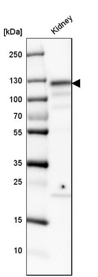
Anti-ACE2 Antibody
Polyclonal Antibody against HUMAN ACE2
0.1 mg/ml


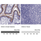
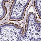
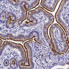
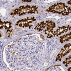
Immunohistochemistry analysis in human small intestine and tonsil tissues using HPA000288 antibody. Corresponding ACE2 RNA-seq data are presented for the same tissues.
Alternative antibodies
Corresponding antigens
With Atlas Antibodies you get

From our facilities in Stockholm, Sweden we develop, manufacture and distribute highly advanced reagents to the Life Sciences community worldwide.

Our products are available to customers worldwide. From most locations, you can order our products from Atlas Antibodies. Please see more information about how to order here.

Learn how we validate our antibodies, how we secure their reproducibility, and why we apply enhanced validation. Our antibodies are validated in IHC, ICC-IF, and WB.